

Carotid Ultrasound Imaging
Ultrasound Imaging, also called ultrasound scanning or sonography, involves exposing part of the body to high-frequency sound waves to produce pictures of the inside of the body. Ultrasound exams do not use ionizing radiation (as used in X-rays). Because ultrasound images are captured in real-time, they can show the structure and movement of the body’s internal organs, as well as blood flowing through blood vessels. Ultrasound imaging is a noninvasive medical test that helps physicians diagnose and treat medical conditions.
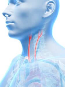
An ultrasound of the body’s two carotid arteries, which are located on each side of the neck and carry blood from the heart to the brain, provide detailed pictures of these blood vessels. A doppler ultrasound study may be part of a carotid ultrasound examination. doppler ultrasound is a special ultrasound technique that evaluates blood as it flows through a blood vessel, including the body’s major arteries and veins in the abdomen, arms, legs and neck.
Benefits of the Procedure
- Most ultrasound scanning is noninvasive (no needles or injections) and is usually painless.
- Ultrasound is widely available, easy-to-use and less expensive than other imaging methods.
- Ultrasound imaging uses no ionizing radiation.
- Ultrasound scanning gives a clear picture of soft tissues that do not show up well on X-ray images.
- Ultrasound causes no health problems and may be repeated as often as is necessary.
- If a carotid ultrasound exam shows narrowing of one or both carotid arteries, treatment can be taken to restore the free flow of blood to the brain. Many strokes are prevented as a result.
The carotid ultrasound is most frequently performed to detect narrowing, or stenosis, of the carotid artery, a condition that substantially increases the risk of stroke. The major goal of carotid ultrasound is to screen patients for blockage or narrowing of their carotid arteries, which if present may increase their risk of having a stroke. Once the diagnosis is made a comprehensive treatment may be initiated. It may also be performed if a patient has high blood pressure or a carotid bruit (pronounced brU-E)—an abnormal sound in the neck that is heard with the stethoscope.
Other risk factors calling for a carotid ultrasound are:
- Advanced age
- Diabetes
- Elevated blood cholesterol
- Family history of stroke or heart disease
A carotid ultrasound is also performed to:
- Locate a hematoma, a collection of clotted blood that may slow and eventually stop blood flow.
- Detect dissection of the carotid artery, a split between layers of the artery wall that may lead to obstruction of blood flow or a weakening of the wall of the artery.
- Check the state of the carotid artery after surgery to restore normal blood flow.
- Verify the position of a metal stent placed to maintain carotid blood flow.
Doppler ultrasound images can help the physician to see and evaluate:
- Blockages to blood flow (such as clots)
- Narrowing of vessels (which may be caused by plaque)
- Tumors and congenital malformation
Preparation
- Wear comfortable, loose-fitting clothing for your ultrasound exam.
- Remove all clothing and jewelry in the area to be examined.
- Wear a loose-fitting, open necked shirt or blouse.
- No other preparation is required.
About the Procedure
Ultrasound imaging is based on the same principles involved in the sonar used by bats, ships and fishermen. When a sound wave strikes an object, it bounces back, or echoes. By measuring these echo waves it is possible to determine how far away the object is and its size, shape, and consistency (whether the object is solid, filled with fluid, or both). In medicine, ultrasound is used to detect changes in appearance of organs, tissues, and vessels or detect abnormal masses, such as tumors. In an ultrasound examination, a transducer both sends the sound waves and records the echoing waves. When the transducer is pressed against the skin, it directs small pulses of inaudible, high-frequency sound waves into the body. As the sound waves bounce off of internal organs, fluids and tissues, the sensitive microphone in the transducer records tiny changes in the sound’s pitch and direction. These signature waves are instantly measured and displayed by a computer, which in turn creates a real-time picture on the monitor. One or more frames of the moving pictures are typically captured as still images.
Doppler ultrasound, a special application of ultrasound, measures the direction and speed of blood cells as they move through vessels. The movement of blood cells causes a change in pitch of the reflected sound waves (called the doppler effect). A computer collects and processes the sounds and creates graphs or color pictures that represent the flow of blood through the blood vessels.
For most ultrasound exams, the patient is positioned lying face-up on an examination table that can be tilted or moved. A clear gel is applied to the area of the body being studied to help the transducer make secure contact with the body and eliminate air pockets between the transducer and the skin. The sonographer (ultrasound technologist) or radiologist then presses the transducer firmly against the skin and sweeps it back and forth over the area of interest.
Doppler sonography is performed using the same transducer. When the examination is complete, the patient may be asked to dress and wait while the ultrasound images are reviewed. However, the sonographer or radiologist is often able to review the ultrasound images in real-time as they are acquired and the patient can be released immediately. This ultrasound examination is usually completed within 30 minutes. After an ultrasound exam, you should be able to resume your normal activities.
Call to Schedule an Appointment
Alpharetta Internal Medicine Office
1380 Upper Hembree Rd.
Roswell, GA 30076
Cumming Internal Medicine Office
950 Sanders Rd
Cumming, GA 30041