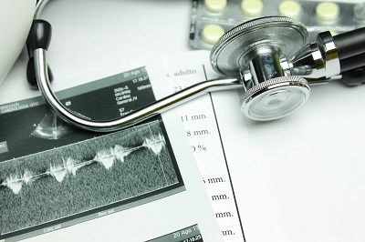
Echocardiography

Echocardiography or ultrasound of the heart is one of the most common procedures in cardiology. This technology involves sending sound waves into the chest through a probe placed on the skin of the chest wall and generating an image with the returning sound waves that reflect off the structures within the heart. These sound waves are high frequency and not audible by human ears. The procedure is virtually harmless, involves no X-rays, and poses little or no known risk to a patient other than the discomfort when the probe is gently pressed against the skin of the chest.
Types of Echocardiography
- One-Dimensional: One-Dimensional or M-mode echocardiography is one beam of ultrasound directed toward the heart. Doctors most often use M-mode echocardiography to see just the left side (or main pumping chamber) of your heart.
- Two-Dimensional: Two-Dimensional echocardiography produces a broader moving picture of your heart. Two-dimensional echocardiography is one of the most important diagnostic tools for doctors.
- Doppler Echocardiography: Doppler echocardiography measures blood flowing through the arteries and shows the pattern of flow through the heart.
About the Procedure
No special preparation is needed before you have an echocardiogram. During the test, you will lie on an examination table. A sonographer will place small metal disks called electrodes on your chest. These electrodes have wires called leads, which hook up to an electrocardiogram machine. This machine will monitor your heart rhythm during the test.
Next, the sonographer will put a thick gel on your chest. The gel may feel cold, but it does not harm your skin. Then, the sonographer will use the transducer to send and receive the sound waves. The transducer will be placed directly on the left side of your chest, above your heart. The sonographer will press firmly as he or she moves the transducer across your chest. You may be asked to breathe in or out or to briefly hold your breath during the test. But, for most of the test, you will lie still.
Call to Schedule an Appointment
Alpharetta Internal Medicine Office
1380 Upper Hembree Rd.
Roswell, GA 30076
Cumming Internal Medicine Office
950 Sanders Rd
Cumming, GA 30041

